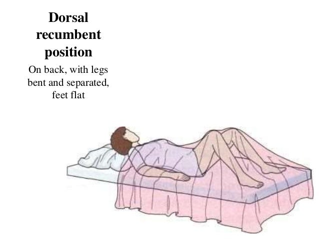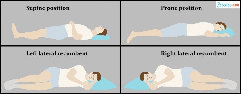
2, 6 The AG measurement with the least intra- and inter-observer variability is the height of the caudal pole in the longitudinal image plane, acquired from dorsal recumbency. 8, 9ĪG sonography is observer-dependent and acquiring measurements is challenging. 10– 12 The effects of sex and age on AG size are variable 4, 6, 7, 9, 10 whereas the influence of body weight leads to the current recommendation to assess AG size in relation to the dog’s weight. 4– 9 These objective measurements may be a value derived from normal dogs 7– 9 or a cut-off separating normal dogs from dogs with PDH. The ultrasound appearance of the normal canine AG was first described in 1990 3 with subsequent definitions of objective measurements. 1 Specifically in PDH, AG ultrasound determines if the AGs are normal size or if bilateral symmetrical hyperplasia exists. Once a diagnosis of HAC is established then AG ultrasound and endocrine differentiating tests assist in the differentiation between pituitary-dependent hyperadrenocorticism (PDH) and an adrenal tumor. Keywords: adrenal glands, ultrasonography, normal, dog, recumbencyĪdrenal gland (AG) ultrasound is part of the assessment of dogs with potential hyperadrenocorticism (HAC), and is interpreted with history, clinical signs, physical examination, routine laboratory tests and endocrine screening tests. This finding suggests that the CPT from the longitudinal image plane is the most reliable measurement in terms of agreement between dorsal and lateral recumbency in dogs with non-AG illness. This study demonstrates that there is some difference in the measurements acquired in dorsal compared with lateral recumbency however, the difference is small for the CPT from the longitudinal plane. Agreement between lateral and dorsal recumbency was poorer for the measurements derived from the transverse image plane and poorest for measurements of AG length in the longitudinal plane. The measurement with the best agreement between the dorsal and lateral recumbency was the caudal pole thickness (CPT) from the longitudinal image plane.

The level and limits of agreement between the dorsal and lateral recumbency for each of the measurements were determined using the Bland–Altman analysis. AG length, height and width measurements made in the longitudinal image plane, and height and width measurements from the transverse image plane were assessed. This prospective study, performed in dogs with non-adrenal illness, compared ultrasonographic AG measurements made in dogs placed in dorsal recumbency with those made in left or right lateral recumbency. The effect of the dogs’ recumbency position on the accuracy of AG measurement acquisition is not known.

#Dorsal recumbent position trial
Poor atitude – if examining fingers will meet an obstruction on the same side as fetal back (hyperextended head)Also palpates infant’s anteroposterior position.Anne Marie Rose, 1 Thurid Johnstone, 1,2 Sue Finch, 3 Cathy Beck 1ġU-Vet Animal Hospital Werribee, The University of Melbourne, 2TRACTS Translational Research and Clinical Trial Study Group, The University of Melbourne, 3Statistical Consulting Centre, School of Mathematics and Statistics, The University of Melbourne, Melbourne, VIC, AustraliaĪbstract: Abdominal ultrasound is frequently used to assess the canine adrenal gland (AG) and subjective and objective features of normal AGs have been described. Good attitude – if brow correspond to the side (2nd maneuver) that contained the elbows and knees. To determine the degree of flexion of fetal head.To determine attitude or habitus.įacing foot part of the woman, palpate fetal head pressing downward about 2 inches above the inguinal ligament. The presenting part is notengaged if it is not movable.It is not yet engaged if it is still movable. Using thumb and finger, grasp the lower portion of the abdomen above symphisis pubis, press in slightly and make gentle movements from side to side. To determine engagement of presenting part. Use gentle but deep pressure.įetal back is smooth, hard, and resistant surface Knees and elbows of fetus feel with a number of angular nodulation One hand is used to steady the uterus on one side of the abdomen while the other hand moves slightly on a circular motion from top to the lower segment of the uterus to feel for the fetal back and small fetal parts.

Breech is less well defined that moves only in conjunction with the body. Head is more firm, hard and round that moves independently of the body. Using both hands, feel for the fetal part lying in the fundus. To determine fetal part lying in the fundus.To determine presentation.


 0 kommentar(er)
